

Mostra el registre d'ítem simple
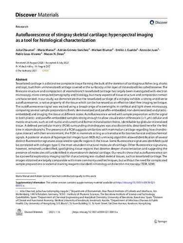
Autofluorescence of stingray skeletal cartilage: hyperspectral imaging as a tool for histological characterization
| dc.contributor.author | de Juan, Ana |
| dc.contributor.author | Dean, Mason |
| dc.contributor.author | Gualda Manzano, Emilio José |
| dc.contributor.author | Marsal, Maria |
| dc.contributor.author | Loza Álvarez, Pablo |
| dc.contributor.author | Chaumel, Júlia |
| dc.contributor.author | Gómez Sánchez, Adrián |
| dc.contributor.author | Blumer, Michael |
| dc.contributor.other | Universitat Politècnica de Catalunya. Departament d'Enginyeria Agroalimentària i Biotecnologia |
| dc.date.accessioned | 2021-11-30T07:03:04Z |
| dc.date.available | 2021-11-30T07:03:04Z |
| dc.date.issued | 2021-12 |
| dc.identifier.citation | De Juan, A. [et al.]. Autofluorescence of stingray skeletal cartilage: hyperspectral imaging as a tool for histological characterization. Springer Science and Business Media LLC, 2021. DOI 10.1007/s43939-021-00015-x. |
| dc.identifier.issn | 2730-7727 |
| dc.identifier.uri | http://hdl.handle.net/2117/357256 |
| dc.description.abstract | Tessellated cartilage is a distinctive composite tissue forming the bulk of the skeleton of cartilaginous fishes (e.g. sharks and rays), built from unmineralized cartilage covered at the surface by a thin layer of mineralized tiles called tesserae. The finescale structure and composition of elasmobranch tessellated cartilage has largely been investigated with electron microscopy, micro-computed tomography and histology, but many aspects of tissue structure and composition remain uncharacterized. In our study, we demonstrate that the tessellated cartilage of a stingray exhibits a strong and diverse autofluorescence, a native property of the tissue which can be harnessed as an effective label-free imaging technique. The autofluorescence signal was excited using a broad range of wavelengths in confocal and light sheet microscopy, comparing several sample preparations (fresh; demineralized and paraffin-embedded; non-demineralized and plastic-embedded) and imaging the tissue at different scales. Autofluorescence varied with sample preparation with the signal in both plastic- and paraffin-embedded samples strong enough to allow visualization of finescale (=¿1 µm) cellular and matrix structures, such as cell nuclei and current and former mineralization fronts, identifiable by globular mineralized tissue. A defined pericellular matrix (PCM) surrounding chondrocytes was also discernible, described here for the first time in elasmobranchs. The presence of a PCM suggests similarities with mammalian cartilage regarding how chondrocytes interact with their environment, the PCM in mammals acting as a transducer for biomechanical and biochemical signals. A posterior analysis of hyperspectral images by an MCR-ALS unmixing algorithm allowed identification of several distinct fluorescence signatures associated to specific regions in the tissue. Some fluorescence signatures identified could be correlated with collagen type II, the most abundant structural molecule of cartilage. Other fluorescence signatures, however, remained unidentified, spotlighting tissue regions that deserve deeper characterization and suggesting the presence of molecules still unidentified in elasmobranch skeletal cartilage. Our results show that autofluorescence can be a powerful exploratory imaging tool for characterizing less-studied skeletal tissues, such as tessellated cartilage. The images obtained are largely comparable with more commonly used techniques, but without the need for complicated sample preparations or external staining reagents standard in histology and electron microscopy (TEM, SEM). |
| dc.format.extent | 28 p. |
| dc.language.iso | eng |
| dc.publisher | Springer Science and Business Media LLC |
| dc.rights | Attribution-NonCommercial-NoDerivs 3.0 Spain |
| dc.rights.uri | http://creativecommons.org/licenses/by-nc-nd/3.0/es/ |
| dc.subject | Àrees temàtiques de la UPC::Física::Física de l'estat sòlid::Luminiscència |
| dc.subject.lcsh | Confocal microscopy |
| dc.subject.other | Tessellated cartilage |
| dc.subject.other | Autofluorescence |
| dc.subject.other | Hyperspectral imaging |
| dc.subject.other | Confocal microscopy |
| dc.subject.other | Light sheet fluorescence microscopy |
| dc.title | Autofluorescence of stingray skeletal cartilage: hyperspectral imaging as a tool for histological characterization |
| dc.type | Audiovisual |
| dc.subject.lemac | Fluorescència |
| dc.identifier.doi | 10.1007/s43939-021-00015-x |
| dc.relation.publisherversion | https://link.springer.com/article/10.1007/s43939-021-00015-x |
| dc.rights.access | Open Access |
| local.identifier.drac | 32001992 |
| dc.description.version | Postprint (published version) |
| local.citation.author | de Juan, A.; Dean, M.; Gualda Manzano, Emilio Jose; Marsal, M.; Loza, P.; Chaumel, J.; Gómez, A.; Blumer, M. |
| local.citation.publicationName | Discover Materials |
| local.citation.volume | 1 |
| local.citation.number | 1 |
| local.citation.startingPage | 1 |
| local.citation.endingPage | 28 |
Fitxers d'aquest items
Aquest ítem apareix a les col·leccions següents
-
Audiovisual [1]